Back أكوابورين Arabic Akvaporin BS Aquaporina Catalan Akvaporin Czech Aquaporin Danish Aquaporine German Υδατοπορίνες Greek Acuaporina Spanish Akvaporiin Estonian آکواپورین Persian
| Aquaporin | |||||||||
|---|---|---|---|---|---|---|---|---|---|
 | |||||||||
| Identifiers | |||||||||
| Symbol | Aquaporin | ||||||||
| Pfam | PF00230 | ||||||||
| InterPro | IPR000425 | ||||||||
| PROSITE | PDOC00193 | ||||||||
| SCOP2 | 1fx8 / SCOPe / SUPFAM | ||||||||
| TCDB | 1.A.8 | ||||||||
| OPM superfamily | 7 | ||||||||
| OPM protein | 2zz9 | ||||||||
| |||||||||
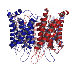
Aquaporins, also called water channels, are channel proteins from a larger family of major intrinsic proteins that form pores in the membrane of biological cells, mainly facilitating transport of water between cells.[1] The cell membranes of a variety of different bacteria, fungi, animal and plant cells contain aquaporins through which water can flow more rapidly into and out of the cell than by diffusing through the phospholipid bilayer.[2] Aquaporins have six membrane-spanning alpha helical domains with both carboxylic and amino terminals on the cytoplasmic side. Two hydrophobic loops contain conserved asparagine–proline–alanine ("NPA motif") which form a barrel surrounding a central pore-like region that contains additional protein density.[3] Because aquaporins are usually always open and are prevalent in just about every cell type, this leads to a misconception that water readily passes through the cell membrane down its concentration gradient. Water can pass through the cell membrane through simple diffusion because it is a small molecule, and through osmosis, in cases where the concentration of water outside of the cell is greater than that of the inside. However, because water is a polar molecule this process of simple diffusion is relatively slow, and in tissues with high water permeability the majority of water passes through aquaporin.[4][5]
The 2003 Nobel Prize in Chemistry was awarded jointly to Peter Agre for the discovery of aquaporins[6] and Roderick MacKinnon for his work on the structure and mechanism of potassium channels.[7]
Genetic defects involving aquaporin genes have been associated with several human diseases including nephrogenic diabetes insipidus and neuromyelitis optica.[8][9][10][11]
- ^ Agre P (2006). "The aquaporin water channels". Proc Am Thorac Soc. 3 (1): 5–13. doi:10.1513/pats.200510-109JH. PMC 2658677. PMID 16493146.
- ^ Cooper G (2009). The Cell: A Molecular Approach. Washington, DC: ASM PRESS. p. 544. ISBN 978-0-87893-300-6.
- ^ Verkman, AS (January 2000). "Structure and function of aquaporin water channels". Am J Physiol Renal Physiol. 278 (1): F13-28. doi:10.1152/ajprenal.2000.278.1.F13. PMID 10644652.
- ^ Cooper, Geoffrey (2000). The Cell (2 ed.). MA: Sinauer Associates. Retrieved 23 April 2020.
- ^ Lodish, Harvey; Berk, Arnold; Zipursky, S. Lawrence (2000). Molecular Cell Biology (4th ed.). New York: W. H. Freeman. ISBN 9781464183393. Retrieved 20 May 2020.
- ^ Knepper MA, Nielsen S (2004). "Peter Agre, 2003 Nobel Prize winner in chemistry". J. Am. Soc. Nephrol. 15 (4): 1093–5. doi:10.1097/01.ASN.0000118814.47663.7D. PMID 15034115.
- ^ "The Nobel Prize in Chemistry 2003". Nobel Foundation. Retrieved 2008-01-23.
- ^ Cite error: The named reference
pmid16087714was invoked but never defined (see the help page). - ^ Cite error: The named reference
pmid16580609was invoked but never defined (see the help page). - ^ Agre P, Kozono D (2003). "Aquaporin water channels: molecular mechanisms for human diseases". FEBS Lett. 555 (1): 72–8. doi:10.1016/S0014-5793(03)01083-4. PMID 14630322. S2CID 35406097.
- ^ Schrier RW (2007). "Aquaporin-related disorders of water homeostasis". Drug News Perspect. 20 (7): 447–53. doi:10.1358/dnp.2007.20.7.1138161. PMID 17992267.
© MMXXIII Rich X Search. We shall prevail. All rights reserved. Rich X Search