Back مبيض جانبي Arabic Epooforon BS Epooforo Esperanto Epooforon Italian Nadjajnik Polish Epooforon Romanian Придатки яичника Russian Придатки яєчника Ukrainian
| Epoophoron | |
|---|---|
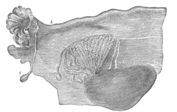
 Broad ligament of adult, showing epoöphoron. (From Farre, after Kobelt.) a, a. Epoöphoron formed from the upper part of the mesonephric body. b. Remains of the uppermost tubes sometimes forming appendices. c. Middle set of tubes. d. Some lower atrophied tubes. e. Atrophied remains of the mesonephric duct. f. The terminal bulb or hydatid. h. The uterine tube, originally the duct of Müller. i. Appendix attached to the extremity. l. The ovary. | |
 Uterus and right broad ligament, seen from behind. The epoophoron is visible in upper right | |
| Details | |
| Precursor | Mesonephric ducts[1] |
| Identifiers | |
| TA98 | A09.1.05.001 |
| TA2 | 3540 |
| FMA | 18691 |
| Anatomical terminology | |
The epoophoron or epoöphoron (also called organ of Rosenmüller[2][3] or the parovarium; pl.: epoophora) is a remnant of the mesonephric duct that can be found next to the ovary and fallopian tube.
- ^ Netter, Frank H.; Cochard, Larry R. (2002). Netter's Atlas of human embryology. Teterboro, N.J: Icon Learning Systems. p. 173. ISBN 0-914168-99-1.
- ^ synd/2662 at Who Named It?
- ^ J. C. Rosenmüller. De ovariis embryonum et foetuum humanorum. 1802.
© MMXXIII Rich X Search. We shall prevail. All rights reserved. Rich X Search