Back Hidroksielapatiet Afrikaans هيدروكسيل أباتيت Arabic Hidroksiapatit BS Hidroxilapatita Catalan Hydroxylapatit Czech Hydroxylapatit German Hidroxiapatita Spanish Hidroxilapatita Basque هیدروکسی آپاتیت Persian Hydroxyapatite French
| Hydroxyapatite | |
|---|---|
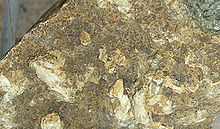
 Hydroxyapatite crystals on matrix | |
| General | |
| Category | Phosphate mineral Apatite group |
| Formula (repeating unit) | Ca5(PO4)3OH |
| IMA symbol | Hap[1] |
| Strunz classification | 8.BN.05 |
| Crystal system | Hexagonal |
| Crystal class | Dipyramidal (6/m) H-M symbol (6/m) |
| Space group | P63/m |
| Unit cell | a = 9.41 Å, c = 6.88 Å; Z = 2 |
| Identification | |
| Formula mass | 502.31 g/mol |
| Color | Colorless, white, gray, yellow, yellowish green |
| Crystal habit | As tabular crystals and as stalagmites, nodules, in crystalline to massive crusts |
| Cleavage | Poor on {0001} and {1010} |
| Fracture | Conchoidal |
| Tenacity | Brittle |
| Mohs scale hardness | 5 |
| Luster | Vitreous to subresinous, earthy |
| Streak | White |
| Diaphaneity | Transparent to translucent |
| Specific gravity | 3.14–3.21 (measured), 3.16 (calculated) |
| Optical properties | Uniaxial (−) |
| Refractive index | nω = 1.651 nε = 1.644 |
| Birefringence | δ = 0.007 |
| References | [2][3][4] |


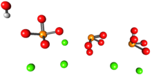
Hydroxyapatite (IMA name: hydroxylapatite[5]) (Hap, HAp, or HA) is a naturally occurring mineral form of calcium apatite with the formula Ca5(PO4)3(OH), often written Ca10(PO4)6(OH)2 to denote that the crystal unit cell comprises two entities.[6] It is the hydroxyl endmember of the complex apatite group. The OH− ion can be replaced by fluoride or chloride, producing fluorapatite or chlorapatite. It crystallizes in the hexagonal crystal system. Pure hydroxyapatite powder is white. Naturally occurring apatites can, however, also have brown, yellow, or green colorations, comparable to the discolorations of dental fluorosis.
Up to 50% by volume and 70% by weight of human bone is a modified form of hydroxyapatite, known as bone mineral.[7] Carbonated calcium-deficient hydroxyapatite is the main mineral of which dental enamel and dentin are composed. Hydroxyapatite crystals are also found in pathological calcifications such as those found in breast tumors,[8] as well as calcifications within the pineal gland (and other structures of the brain) known as corpora arenacea or "brain sand".[9]
- ^ Warr, L.N. (2021). "IMA–CNMNC approved mineral symbols". Mineralogical Magazine. 85 (3): 291–320. Bibcode:2021MinM...85..291W. doi:10.1180/mgm.2021.43. S2CID 235729616.
- ^ Hydroxylapatite on Mindat
- ^ Hydroxylapatite on Webmineral
- ^ Anthony, John W.; Bideaux, Richard A.; Bladh, Kenneth W.; Nichols, Monte C., eds. (2000). "Hydroxylapatite". Handbook of Mineralogy (PDF). Vol. IV (Arsenates, Phosphates, Vanadates). Chantilly, VA, US: Mineralogical Society of America. ISBN 978-0962209734. Archived (PDF) from the original on 2018-09-29. Retrieved 2010-08-29.
- ^ "The official IMA-CNMNC List of Mineral Names". International Mineralogical Association: COMMISSION ON NEW MINERALS, NOMENCLATURE AND CLASSIFICATION. Retrieved 24 August 2023.
- ^ Singh, Anamika; Tiwari, Atul; Bajpai, Jaya; Bajpai, Anil K. (2018-01-01), Tiwari, Atul (ed.), "3 – Polymer-Based Antimicrobial Coatings as Potential Biomaterials: From Action to Application", Handbook of Antimicrobial Coatings, Elsevier, pp. 27–61, doi:10.1016/b978-0-12-811982-2.00003-2, ISBN 978-0-12-811982-2, retrieved 2020-11-18
- ^
Junqueira, Luiz Carlos; José Carneiro (2003). Foltin, Janet; Lebowitz, Harriet; Boyle, Peter J. (eds.). Basic Histology, Text & Atlas (10th ed.). McGraw-Hill Companies. p. 144. ISBN 978-0-07-137829-1.
Inorganic matter represents about 50% of the dry weight of bone ... crystals show imperfections and are not identical to the hydroxylapatite found in the rock minerals
- ^ Haka, Abigail S.; Shafer-Peltier, Karen E.; Fitzmaurice, Maryann; Crowe, Joseph; Dasari, Ramachandra R.; Feld, Michael S. (2002-09-15). "Identifying microcalcifications in benign and malignant breast lesions by probing differences in their chemical composition using Raman spectroscopy". Cancer Research. 62 (18): 5375–5380. ISSN 0008-5472. PMID 12235010.
- ^ Angervall, Lennart; Berger, Sven; Röckert, Hans (2009). "A Microradiographic and X-Ray Crystallographic Study of Calcium in the Pineal Body and in Intracranial Tumours". Acta Pathologica et Microbiologica Scandinavica. 44 (2): 113–19. doi:10.1111/j.1699-0463.1958.tb01060.x. PMID 13594470.
© MMXXIII Rich X Search. We shall prevail. All rights reserved. Rich X Search