Back S'ueb ACE Long Afrikaans Lunge ALS ሳምባ Amharic Polmón AN رئة Arabic ܪܐܬܐ ARC হাঁওফাঁও Assamese Pulmón AST Opan ATJ
| Lung | |
|---|---|
 Diagram of the human lungs with the respiratory tract visible, and different colours for each lobe | |
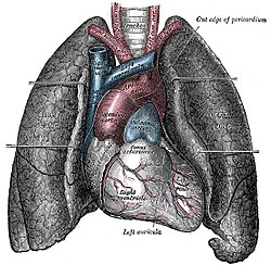
 The human lungs flank the heart and great vessels in the chest cavity. | |
| Details | |
| System | Respiratory system |
| Artery | Pulmonary Artery |
| Vein | Pulmonary Vein |
| Identifiers | |
| Latin | pulmo |
| Greek | πνεύμων (pneumon) |
| MeSH | D008168 |
| TA98 | A06.5.01.001 |
| TA2 | 3265 |
| Anatomical terminology | |
The lungs are the main organs of the respiratory system in most terrestrial animals, including all tetrapod vertebrates and a small number of amphibious fish (lungfish and bichirs), pulmonate gastropods (land snails and slugs, which have analogous pallial lungs), and some arachnids (tetrapulmonates such as spiders and scorpions, which have book lungs). Their function is to conduct gas exchange by extracting oxygen from the air into the bloodstream via diffusion, and to release carbon dioxide from the bloodstream out into the atmosphere, a process also known as respiration. This article primarily concerns with the lungs of tetrapods (particularly those of humans), which are paired and located on either side of the heart, occupying most of the volume of the thoracic cavity, and are homologous to the swim bladders in ray-finned fish.
The movements of air in and out of the lungs is called ventilation or breathing, which is driven by different muscular systems in different species. Amniotes like mammals, reptiles and birds use different dedicated respiratory muscles to facilitate breathing, while in primitive tetrapods, air was driven into the lungs by the pharyngeal muscles via buccal pumping, a mechanism still seen in amphibians. In humans, the main muscles of respiration that drives breathing are the diaphragm and intercostal muscles, while other core and limb muscles might also be recruited as accessory muscles in situations of respiratory distress. The lungs also provide airflow that makes vocalization (including human speech) possible.
Human lungs, like other tetrapods, are paired with one on the left and one on the right. Due to the leftward rotation of the heart, the right lung is bigger and heavier than the left, and the two lungs together weigh approximately 1.3 kilograms (2.9 lb). The lungs are part of the lower respiratory tract that begins at the trachea and branches into the bronchi and bronchioles, and which receive fresh air inhaled (breathed in) via the conducting zone. The conducting zone ends at the terminal bronchioles, which divide into the respiratory bronchioles of the respiratory zone and further divide into alveolar ducts that give rise to the alveolar sacs that contain the alveoli, where majority of gas exchange takes place. Alveoli are also sparsely present on the walls of the respiratory bronchioles and alveolar ducts. Together, the lungs contain approximately 2,400 kilometres (1,500 mi) of airways and 300 to 500 million alveoli. Each lung is enveloped by a serous membranes called pleurae, which also overlays the inside surface of the rib cage; the two membranes (called the visceral and parietal pleurae, respectively) form an enclosing sac known as the pleural cavity, which contains a lubricating film of serous fluid (pleural fluid) that separates the two pleurae and reduces the friction of sliding movements between them, allowing for easier expansion of the lungs during breathing. The visceral pleura also invaginates into each lung as fissures, which divides the lung into independent sections called lobes. The right lung typically has three lobes, and the left has two. The lobes are further divided into bronchopulmonary segments and pulmonary lobules.
The lungs have two unique blood supplies: the pulmonary circulation, which receives deoxygenated blood from the right heart via the pulmonary arteries, exchanges oxygen and carbon dioxide across the alveolar–capillary barrier, before returning the re-oxygenated blood to the left heart via the pulmonary veins for pumping to the rest of the body; and the bronchial circulation, which is part of the systemic circulation that provides a separate supply of oxygenated blood to the tissue of the lungs.[1][2]
The lung can be affected by a number of respiratory diseases, including pneumonia, pulmonary fibrosis and lung cancer. Chronic obstructive pulmonary disease includes chronic bronchitis and emphysema, and are commonly related to smoking or exposure to air pollutants. A number of occupational lung diseases can be caused by substances such as coal dust, asbestos fibres and crystalline silica dust. Diseases such as acute bronchitis and asthma can also affect lung function, although such conditions are technically airway diseases rather than lung diseases. Medical terms related to the lung often begin with pulmo-, from the Latin pulmonarius (meaning "of the lungs") as in pulmonology, or with pneumo- (from Greek πνεύμων, meaning "lung") as in pneumonia.
In embryonic development, the lungs begin to develop as an outpouching of the foregut, a tube which goes on to form the upper part of the digestive system. When the lungs are formed the fetus is held in the fluid-filled amniotic sac and so they do not function to breathe. Blood is also diverted from the lungs through the ductus arteriosus. At birth, air begins to pass through the lungs, and the diversionary duct closes, so that the lungs can begin to respire. The lungs only fully develop in early childhood.
- ^ Association, American Lung. "How Lungs Work". www.lung.org. Retrieved 2023-11-18.
- ^ Tucker, William D.; Weber, Carly; Burns, Bracken (2023), "Anatomy, Thorax, Heart Pulmonary Arteries", StatPearls, Treasure Island (FL): StatPearls Publishing, PMID 30521233, retrieved 2023-11-18
© MMXXIII Rich X Search. We shall prevail. All rights reserved. Rich X Search