Back بصلة شمية Arabic Njušni režanj BS Bulb olfactori Catalan Čichový bulbus Czech Riechkolben German Bulbo olfatorio Spanish Haistmissibul Estonian Usaimen-erraboil Basque پیاز بویایی Persian Bulbe olfactif French
| Olfactory bulb | |
|---|---|
 Human brain seen from below. Vesalius' Fabrica, 1543. Olfactory bulbs and olfactory tracts outlined in red | |
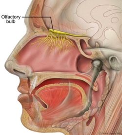
 Sagittal section of human head. | |
| Details | |
| System | Olfactory |
| Identifiers | |
| Latin | bulbus olfactorius |
| MeSH | D009830 |
| NeuroNames | 279 |
| NeuroLex ID | birnlex_1137 |
| TA98 | A14.1.09.429 |
| TA2 | 5538 |
| FMA | 77624 |
| Anatomical terms of neuroanatomy | |

Blue – Glomerular layer;
Red – External Plexiform and Mitral cell layer;
Green – Internal Plexiform and Granule cell layer.
Top of image is dorsal aspect, right of image is lateral aspect. Scale, ventral to dorsal, is approximately 2mm.
The olfactory bulb (Latin: bulbus olfactorius) is a neural structure of the vertebrate forebrain involved in olfaction, the sense of smell. It sends olfactory information to be further processed in the amygdala, the orbitofrontal cortex (OFC) and the hippocampus where it plays a role in emotion, memory and learning. The bulb is divided into two distinct structures: the main olfactory bulb and the accessory olfactory bulb. The main olfactory bulb connects to the amygdala via the piriform cortex of the primary olfactory cortex and directly projects from the main olfactory bulb to specific amygdala areas. The accessory olfactory bulb resides on the dorsal-posterior region of the main olfactory bulb and forms a parallel pathway. Destruction of the olfactory bulb results in ipsilateral anosmia, while irritative lesions of the uncus can result in olfactory and gustatory hallucinations.

© MMXXIII Rich X Search. We shall prevail. All rights reserved. Rich X Search