Back اسطوانة بولية Arabic Zylinder (Urin) German Ίζημα ούρων Greek Cilindros urinarios Spanish Cilindro urinario Italian 尿円柱 Japanese Wałeczki Polish 管型 Chinese
This article needs additional citations for verification. (March 2023) |

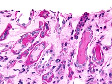
Urinary casts are microscopic cylindrical structures produced by the kidney and present in the urine in certain disease states. They form in the distal convoluted tubule and collecting ducts of nephrons, then dislodge and pass into the urine, where they can be detected by microscopy.
They form via precipitation of Tamm–Horsfall mucoprotein, which is secreted by renal tubule cells, and sometimes also by albumin in conditions of proteinuria. Cast formation is pronounced in environments favoring protein denaturation and precipitation (low flow, concentrated salts, low pH). Tamm–Horsfall protein is particularly susceptible to precipitation in these conditions.
Casts were first described by Henry Bence Jones (1813–1873).[1]
As reflected in their cylindrical form, casts are generated in the small distal convoluted tubules and collecting ducts of the kidney, and generally maintain their shape and composition as they pass through the urinary system. Although the most common forms are benign, others indicate disease. All rely on the inclusion or adhesion of various elements on a mucoprotein base—the hyaline cast. "Cast" itself merely describes the shape, so an adjective is added to describe the composition of the cast. Various casts found in urine sediment may be classified as:
- ^ Louis Rosenfeld, Four Centuries of Clinical Chemistry, p.50, Gordon & Breach Science, 1999 ISBN 90-5699-645-2.
© MMXXIII Rich X Search. We shall prevail. All rights reserved. Rich X Search