Back خلية كأسية Arabic Bechazej BAR Cèl·lula caliciforme Catalan Pohárková buňka Czech Becherzelle German Célula caliciforme Spanish سلول جامی Persian Pikarisolu Finnish Cellule caliciforme French Célula caliciforme Galician
| Goblet cell | |
|---|---|
 Schematic illustration of a goblet cell in close up, illustrating different internal structures of the cell. | |
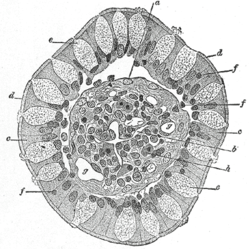
 Transverse section of a villus, from the human intestine. X 350. a. Basement membrane, here somewhat shrunken away from the epithelium. b. Lacteal. c. Columnar epithelium. d. Its striated border. e. Goblet cells. f. Leucocytes in epithelium. f’. Leucocytes below epithelium. g. Blood vessels. h. Muscle cells cut across. | |
| Details | |
| System | Respiratory system |
| Shape | Simple columnar |
| Function | Mucin-producing epithelial cells |
| Identifiers | |
| Latin | exocrimohsinoctus caliciformis |
| MeSH | D020397 |
| TH | H3.04.03.0.00009, H3.04.03.0.00016, H3.05.00.0.00006 |
| FMA | 13148 |
| Anatomical terms of microanatomy | |
Goblet cells are simple columnar epithelial cells that secrete gel-forming mucins, like mucin 2 in the lower gastrointestinal tract, and mucin 5AC in the respiratory tract.[1] The goblet cells mainly use the merocrine method of secretion, secreting vesicles into a duct, but may use apocrine methods, budding off their secretions, when under stress.[2] The term goblet refers to the cell's goblet-like shape. The apical portion is shaped like a cup, as it is distended by abundant mucus laden granules; its basal portion lacks these granules and is shaped like a stem.
The goblet cell is highly polarized with the nucleus and other organelles concentrated at the base of the cell and secretory granules containing mucin, at the apical surface.[1] The apical plasma membrane projects short microvilli to give an increased surface area for secretion.[3]
Goblet cells are typically found in the respiratory, reproductive and lower gastrointestinal tract and are surrounded by other columnar cells.[1] Biased differentiation of airway basal cells in the respiratory epithelium, into goblet cells plays a key role in the excessive mucus production, known as mucus hypersecretion seen in many respiratory diseases, including chronic bronchitis, and asthma.[4][5]
- ^ a b c Hodges, R.R.; Dartt, D.A. (2010). "Conjunctival Goblet Cells". Encyclopedia of the Eye. pp. 369–376. doi:10.1016/b978-0-12-374203-2.00053-1. ISBN 9780123742032.
- ^ Lohmann-Matthes, M-L.; Steinmüller, C.; Franke-Ullmann, G. (1994). "Pulmonary macrophages". European Respiratory Journal. 7 (9): 1678–1689. doi:10.1183/09031936.94.07091678. PMID 7995399.
- ^ Saladin, K (2012). Anatomy & physiology : the unity of form and function (6th ed.). McGraw-Hill. pp. 88–89. ISBN 9780073378251.
- ^ Ohar, JA; Donohue, JF; Spangenthal, S (23 October 2019). "The Role of Guaifenesin in the Management of Chronic Mucus Hypersecretion Associated with Stable Chronic Bronchitis: A Comprehensive Review". Chronic Obstructive Pulmonary Diseases. 6 (4): 341–349. doi:10.15326/jcopdf.6.4.2019.0139. PMC 7006698. PMID 31647856.
- ^ Evans, CM; Kim, K; Tuvim, MJ; Dickey, BF (January 2009). "Mucus hypersecretion in asthma: causes and effects". Current Opinion in Pulmonary Medicine. 15 (1): 4–11. doi:10.1097/MCP.0b013e32831da8d3. PMC 2709596. PMID 19077699.
© MMXXIII Rich X Search. We shall prevail. All rights reserved. Rich X Search