Back بكتيريا إيجابية الغرام Arabic গ্রাম পজিটিভ ব্যাকটেরিয়া Bengali/Bangla Gram-Ya Breton Gram-pozitivna bakterija BS Bacteri grampositiu Catalan Grampozitivní bakterie Czech Gram-positive bakterier Danish Gram-pozitivaj bakterioj Esperanto Bacteria grampositiva Spanish Grampositiivsed bakterid Estonian


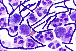
In bacteriology, gram-positive bacteria are bacteria that give a positive result in the Gram stain test, which is traditionally used to quickly classify bacteria into two broad categories according to their type of cell wall.
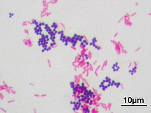
The Gram stain is used by microbiologists to place bacteria into two main categories, Gram-positive (+) and Gram-negative (-). Gram-positive bacteria have a thick layer of peptidoglycan within the cell wall, and Gram-negative bacteria have a thin layer of peptidoglycan.
Gram-positive bacteria take up the crystal violet stain used in the test, and then appear to be purple-coloured when seen through an optical microscope. This is because the thick layer of peptidoglycan in the bacterial cell wall retains the stain after it is washed away from the rest of the sample, in the decolorization stage of the test.
Conversely, gram-negative bacteria cannot retain the violet stain after the decolorization step; alcohol used in this stage degrades the outer membrane of gram-negative cells, making the cell wall more porous and incapable of retaining the crystal violet stain. Their peptidoglycan layer is much thinner and sandwiched between an inner cell membrane and a bacterial outer membrane, causing them to take up the counterstain (safranin or fuchsine) and appear red or pink.
Despite their thicker peptidoglycan layer, gram-positive bacteria are more receptive to certain cell wall–targeting antibiotics than gram-negative bacteria, due to the absence of the outer membrane.[1]
- ^ Basic Biology (18 March 2016). "Bacteria".
© MMXXIII Rich X Search. We shall prevail. All rights reserved. Rich X Search