Back عضلة موسعة للحدقة Arabic Mišić dilatator zjenice BS Múscul dilatador de la pupil·la Catalan Musculus dilatator pupillae German Músculo dilatador del iris Spanish ماهیچه گشادکننده عنبیه Persian Muscle dilatateur de la pupille French Mišić dilatator zjenice Croatian Musculus dilatator pupillae Hungarian Muscolo dilatatore dell'iride Italian
| Iris dilator muscle | |
|---|---|
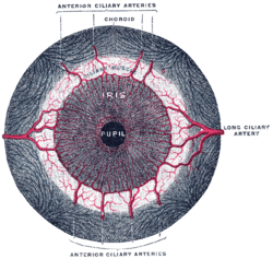
 Iris, front view. (Muscle visible but not labeled.) | |
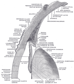
 The upper half of a sagittal section through the front of the eyeball. (Iris dilator muscle is NOT labeled and not to be confused with "Radiating fibers" labeled near center, which are part of the ciliary muscle.) | |
| Details | |
| Origin | Outer margins of iris[1] |
| Insertion | Inner margins of iris[1] |
| Nerve | Long ciliary nerves (sympathetics) |
| Actions | Dilates pupil |
| Antagonist | Iris sphincter muscle |
| Identifiers | |
| Latin | musculus dilatator pupillae |
| TA98 | A15.2.03.030 |
| TA2 | 6763 |
| FMA | 49158 |
| Anatomical terms of muscle | |
The iris dilator muscle (pupil dilator muscle, pupillary dilator, radial muscle of iris, radiating fibers), is a smooth muscle[2] of the eye, running radially in the iris and therefore fit as a dilator. The pupillary dilator consists of a spokelike arrangement of modified contractile cells called myoepithelial cells. These cells are stimulated by the sympathetic nervous system.[3] When stimulated, the cells contract, widening the pupil and allowing more light to enter the eye.
- ^ a b Gest, Thomas R; Burkel, William E. (2000). "Anatomy Tables – Eye". Medical Gross Anatomy. University of Michigan Medical School. Archived from the original on 2010-05-26.
{{cite web}}: CS1 maint: unfit URL (link) - ^ Pilar, G; Nuñez, R; McLennan, I. S.; Meriney, S. D. (1987). "Muscarinic and nicotinic synaptic activation of the developing chicken iris". The Journal of Neuroscience. 7 (12): 3813–3826. doi:10.1523/JNEUROSCI.07-12-03813.1987. PMC 6569112. PMID 2826718.
- ^ Saladin, Kenneth (2012). Anatomy and Physiology. McGraw-Hill. pp. 616–617.
© MMXXIII Rich X Search. We shall prevail. All rights reserved. Rich X Search