Back تخطيط مغناطيسية الدماغ Arabic Magnetoencefalografia Catalan Magnetoencefalografie Czech Magnetoenzephalographie German Magnetoencefalografía Spanish مگنتوانسفالوگرافی Persian Magnetoenkefalografia Finnish Magnétoencéphalographie French מגנטואנצפלוגרפיה HE Magnetoenkefalográfia Hungarian
| Magnetoencephalography | |
|---|---|
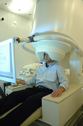
 Person undergoing a MEG | |
| MeSH | D015225 |
Magnetoencephalography (MEG) is a functional neuroimaging technique for mapping brain activity by recording magnetic fields produced by electrical currents occurring naturally in the brain, using very sensitive magnetometers. Arrays of SQUIDs (superconducting quantum interference devices) are currently the most common magnetometer, while the SERF (spin exchange relaxation-free) magnetometer is being investigated for future machines.[1][2] Applications of MEG include basic research into perceptual and cognitive brain processes, localizing regions affected by pathology before surgical removal, determining the function of various parts of the brain, and neurofeedback. This can be applied in a clinical setting to find locations of abnormalities as well as in an experimental setting to simply measure brain activity.[3]
- ^ Hämäläinen M, Hari R, Ilmoniemi RJ, Knuutila J, Lounasmaa OV (1993). "Magnetoencephalography—theory, instrumentation, and applications to noninvasive studies of the working human brain" (PDF). Reviews of Modern Physics. 65 (2): 413–497. Bibcode:1993RvMP...65..413H. doi:10.1103/RevModPhys.65.413. ISSN 0034-6861.
- ^ Boto E, Holmes N, Leggett J, Roberts G, Shah V, Meyer SS, Muñoz LD, Mullinger KJ, Tierney TM (March 2018). "Moving magnetoencephalography towards real-world applications with a wearable system". Nature. 555 (7698): 657–661. Bibcode:2018Natur.555..657B. doi:10.1038/nature26147. ISSN 1476-4687. PMC 6063354. PMID 29562238.
- ^ Carlson NR (2013). Physiology of Behavior. Upper Saddle River, NJ: Pearson Education Inc. pp. 152–153. ISBN 978-0-205-23939-9.
© MMXXIII Rich X Search. We shall prevail. All rights reserved. Rich X Search