Back ظهارة متوسطة Arabic Mezotel Azerbaijani Mesoteli Catalan Mezotel Czech Μεσοθήλιο Greek Mesotelio Spanish مزوتلیوم Persian Mésothélium French Mesotelio Galician Sel mesotelial ID
| Mesothelium | |
|---|---|
 | |
 | |
| Details | |
| Precursor | Somatopleuric mesenchyme |
| Identifiers | |
| Latin | mesothelium |
| TH | H2.00.02.0.02017, H3.04.08.0.00003 |
| FMA | 14074 |
| Anatomical terminology | |
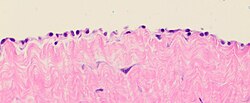
The mesothelium is a membrane composed of simple squamous epithelial cells of mesodermal origin,[2] which forms the lining of several body cavities: the pleura (pleural cavity around the lungs), peritoneum (abdominopelvic cavity including the mesentery, omenta, falciform ligament and the perimetrium) and pericardium (around the heart).
Mesothelial tissue also surrounds the male testis (as the tunica vaginalis) and occasionally the spermatic cord (in a patent processus vaginalis). Mesothelium that covers the internal organs is called visceral mesothelium, while one that covers the surrounding body walls is called the parietal mesothelium. The mesothelium that secretes serous fluid as a main function is also known as a serosa.
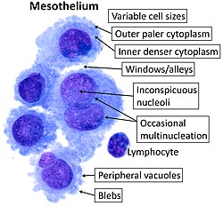
- ^ Image by Mikael Häggström, MD. Sources for mentioned features:
- "Mesothelial cytopathology". Libre Pathology. Retrieved 2022-10-18.
- Shidham VB, Layfield LJ (2021). "Introduction to the second edition of 'Diagnostic Cytopathology of Serous Fluids' as CytoJournal Monograph (CMAS) in Open Access". CytoJournal. 18: 30. doi:10.25259/CMAS_02_01_2021. PMC 8813611. PMID 35126608. - ^ Victor P. Eroschenko (2008). Di Fiore's atlas of histology with functional correlations. Lippincott Williams & Wilkins. pp. 31–. ISBN 978-0-7817-7057-6. Retrieved 28 May 2011.
© MMXXIII Rich X Search. We shall prevail. All rights reserved. Rich X Search