Back صورة مجهرية Arabic Mikrofotoqrafiya Azerbaijani Micrografia Catalan Mikrofotografie Czech Mikrofotografie German Micrografía Spanish عکاسی ریزنگاری Persian Photomicrographie French Fótaimicreagraf Irish सूक्ष्मचित्र Hindi
This article needs additional citations for verification. (September 2014) |


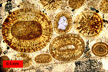
A micrograph or photomicrograph is a photograph or digital image taken through a microscope or similar device to show a magnified image of an object. This is opposed to a macrograph or photomacrograph, an image which is also taken on a microscope but is only slightly magnified, usually less than 10 times. Micrography is the practice or art of using microscopes to make photographs.
A micrograph contains extensive details of microstructure. A wealth of information can be obtained from a simple micrograph like behavior of the material under different conditions, the phases found in the system, failure analysis, grain size estimation, elemental analysis and so on. Micrographs are widely used in all fields of microscopy.
© MMXXIII Rich X Search. We shall prevail. All rights reserved. Rich X Search