| Nasal cartilages | |
|---|---|
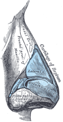
 Cartilages of the nose. Side view. (Greater alar cartilage visible in blue at center right.) | |
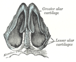
 Cartilages of the nose, seen from below. | |
| Details | |
| Identifiers | |
| Latin | cartilagines nasi |
| MeSH | D055171 |
| TA98 | A06.1.01.006 |
| TA2 | 939 |
| FMA | 59502 |
| Anatomical terminology | |
The nasal cartilages are structures within the nose that provide form and support to the nasal cavity.[1] The nasal cartilages are made up of a flexible material called hyaline cartilage (packed collagen) in the distal portion of the nose.[2] There are five individual cartilages that make up the nasal cavity: septal nasal cartilage, lateral nasal cartilage, major alar cartilage (greater alar cartilage, or cartilage of the aperture), minor alar cartilage (lesser alar cartilage, sesamoid, or accessory cartilage), and vomeronasal cartilage (Jacobson's cartilage).

The nasal cartilages associate with other cartilage structures of the nose or with bones of the facial skeleton. These associations create vent-like structures within the nose so that air can flow from the nasal cavity to the lungs or vice versa. Therefore, the nasal cartilages are structures that aid the body in respiratory functions to intake oxygen or expire carbon dioxide.
Abnormalities or defects in the nasal cartilages affect airflow through the nasal cavity, resulting in respiratory issues. Surgical techniques have been produced to adjust the position or repair the nasal cartilages so that maximal airflow is once again accomplished. Some of these surgical techniques include: septoplasty (restructuring the septal nasal cartilage), upper lateral cartilage repositioning (restructuring the lateral nasal cartilage), and sliding alar cartilage (restructuring the major alar cartilage). Rhinoplasty, which is the surgical reconstruction of the nose, has increased in recent popularity for functional and social purposes.
Other mammals also contain nasal cartilages in order to maintain structure and function for the nasal cavity. The orientation of the nasal cartilages can produce different shapes and sizes of the nostrils and nasal cavities. For the most part, animals contain similar cartilage structures within the nose but vary in the number of different cartilage structures they have. Donkeys, buffalo, and camels have a variety of cartilage structures that are analogous to humans but they all lack septal nasal cartilages. Instead, they have multiple components merging together to form the nasal septum. Even though nasal cartilages differ between species, they all aid in the function of the respiratory system.
- ^ Lang, Johannes (1989). Clinical Anatomy of the Nose, Nasal Cavity and Paranasal Sinuses. Thieme. pp. 10–15. ISBN 978-3-13-738401-4. Retrieved March 11, 2021.
- ^ Krishnan, Yamini; Grodzinsky, Alan J. (2018). "Cartilage Diseases". Matrix Biology. 71–72: 51–69. doi:10.1016/j.matbio.2018.05.005. ISSN 0945-053X. PMC 6146013. PMID 29803938.
© MMXXIII Rich X Search. We shall prevail. All rights reserved. Rich X Search
