Back عيينة (عضو) Arabic Omatidiu AST ওমাটিডিয়াম Bengali/Bangla Ommatidi Catalan Ommatidium German Omatidio Spanish Omatidio Basque ریزچشمک Persian Ommatidi Finnish Ommatidie French


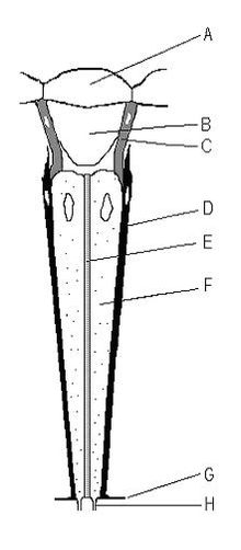
The compound eyes of arthropods like insects, crustaceans and millipedes[1] are composed of units called ommatidia (sg.: ommatidium). An ommatidium contains a cluster of photoreceptor cells surrounded by support cells and pigment cells. The outer part of the ommatidium is overlaid with a transparent cornea. Each ommatidium is innervated by one axon bundle (usually consisting of 6–9 axons, depending on the number of rhabdomeres)[2]: 162 and provides the brain with one picture element. The brain forms an image from these independent picture elements. The number of ommatidia in the eye depends upon the type of arthropod and range from as low as 5 as in the Antarctic isopod Glyptonotus antarcticus,[3] or a handful in the primitive Zygentoma, to around 30,000 in larger Anisoptera dragonflies and some Sphingidae moths.[4]
- ^ Müller CH, Sombke A, Rosenberg J (December 2007). "The fine structure of the eyes of some bristly millipedes (Penicillata, Diplopoda): additional support for the homology of mandibulate ommatidia". Arthropod Structure & Development. 36 (4): 463–76. doi:10.1016/j.asd.2007.09.002. PMID 18089122.
- ^ Land MF, Nilsson DE (2012). Animal Eyes (Second ed.). Oxford University Press. ISBN 978-0-19-958114-6.
- ^ Meyer-Rochow VB (1982). "The divided eye of the isopod Glyptonotus antarcticus: effects of unilateral dark adaptation and temperature elevation". Proceedings of the Royal Society of London. B215 (1201): 433–450. Bibcode:1982RSPSB.215..433M. doi:10.1098/rspb.1982.0052. S2CID 85297324.
- ^ Common IF (1990). Moths of Australia. Brill. p. 15. ISBN 978-90-04-09227-3.
© MMXXIII Rich X Search. We shall prevail. All rights reserved. Rich X Search