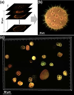
Optical sectioning is the process by which a suitably designed microscope can produce clear images of focal planes deep within a thick sample. This is used to reduce the need for thin sectioning using instruments such as the microtome. Many different techniques for optical sectioning are used and several microscopy techniques are specifically designed to improve the quality of optical sectioning.
Good optical sectioning, often referred to as good depth or z resolution, is popular in modern microscopy as it allows the three-dimensional reconstruction of a sample from images captured at different focal planes.
- ^ Qian, Jia; Lei, Ming; Dan, Dan; Yao, Baoli; Zhou, Xing; Yang, Yanlong; Yan, Shaohui; Min, Junwei; Yu, Xianghua (2015). "Full-color structured illumination optical sectioning microscopy". Scientific Reports. 5: 14513. Bibcode:2015NatSR...514513Q. doi:10.1038/srep14513. PMC 4586488. PMID 26415516.
© MMXXIII Rich X Search. We shall prevail. All rights reserved. Rich X Search
