Back سبيل هرمي Arabic Piramida sistemi Azerbaijani Sistema piramidal Catalan Pyramidebane Danish Pyramidales System German Sistema piramidal Spanish Sistema piramidal Basque راه هرمی Persian Kortikospinaalirata Finnish Faisceau pyramidal French
| Pyramidal tracts | |
|---|---|
 Deep dissection of brain-stem. Lateral view. ("pyramidal tract" visible in red, and "pyramidal decussation" labeled at lower right.) | |
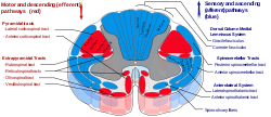
 Spinal cord tracts, with pyramidal tracts labeled at upper left | |
| Details | |
| Decussation | Many fibres in pyramids of medulla oblongata |
| From | Cerebral cortex |
| To | Spinal cord (corticospinal) or brainstem (corticobulbar) |
| Identifiers | |
| Latin | tractus pyramidalis tractus corticospinalis |
| MeSH | D011712 |
| NeuroNames | 1320 |
| NeuroLex ID | birnlex_1464 |
| TA98 | A14.1.04.102 A14.1.06.102 |
| TA2 | 6040 |
| FMA | 72634 |
| Anatomical terms of neuroanatomy | |
The pyramidal tracts include both the corticobulbar tract and the corticospinal tract. These are aggregations of efferent nerve fibers from the upper motor neurons that travel from the cerebral cortex and terminate either in the brainstem (corticobulbar) or spinal cord (corticospinal) and are involved in the control of motor functions of the body.
The corticobulbar tract conducts impulses from the brain to the cranial nerves.[1] These nerves control the muscles of the face and neck and are involved in facial expression, mastication, swallowing, and other motor functions.
The corticospinal tract conducts impulses from the brain to the spinal cord. It is made up of a lateral and anterior tract. The corticospinal tract is involved in voluntary movement. The majority of fibres of the corticospinal tract cross over in the medulla oblongata, resulting in muscles being controlled by the opposite side of the brain. The corticospinal tract contains the axons of the pyramidal cells, the largest of which are the Betz cells, located in the cerebral cortex.
The pyramidal tracts are named because they pass through the pyramids of the medulla oblongata. The corticospinal fibers converge to a point when descending from the internal capsule to the brain stem from multiple directions, giving the impression of an inverted pyramid. Involvement of the pyramidal tract at any level leads to pyramidal signs.
The myelination of the pyramidal fibres is incomplete at birth and gradually progresses in cranio-caudal direction and thereby progressively gaining functionality. Most of the myelination is complete by two years of age and thereafter it progresses very slowly in cranio-caudal direction up to twelve years of age.
- ^ Chapter 9 of "Principles of Physiology" (3rd edition) by Robert M. Berne and Mathew N. Levy. Published by Mosby, Inc. (2000) ISBN 0-323-00813-5.
© MMXXIII Rich X Search. We shall prevail. All rights reserved. Rich X Search