Back Borswerwel Afrikaans فقرة صدرية Arabic Torakal fəqərələr Azerbaijani Mell ar c'hein Breton Grudni pršljenovi BS Vèrtebra dorsal Catalan Кăкăр сыпписем CV Brustwirbel German Θωρακικοί σπόνδυλοι Greek Torakaj vertebroj Esperanto
| Thoracic vertebrae | |
|---|---|
 Position of the thoracic vertebrae (T1–T12) | |
 Animation of T2 | |
| Details | |
| Identifiers | |
| Latin | vertebrae thoracicae |
| MeSH | D013904 |
| TA98 | A02.2.03.001 |
| TA2 | 1059 |
| FMA | 9139 |
| Anatomical terms of bone | |
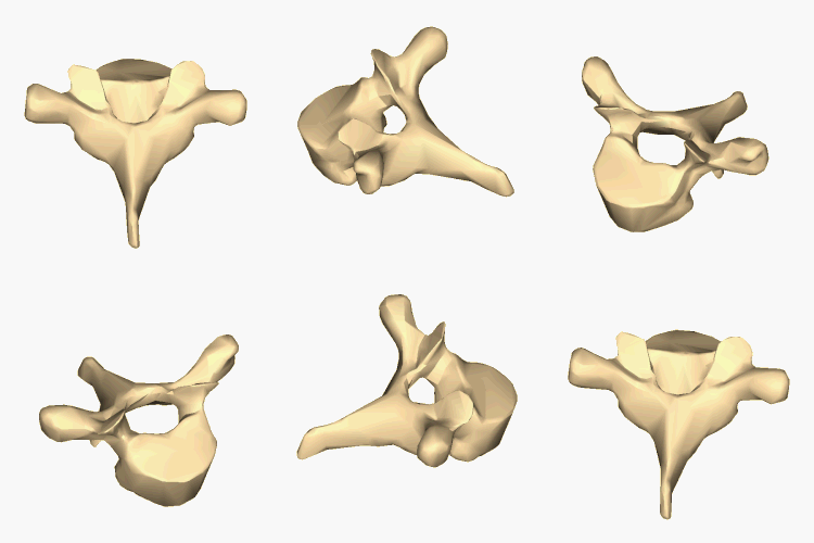
In vertebrates, thoracic vertebrae compose the middle segment of the vertebral column, between the cervical vertebrae and the lumbar vertebrae.[1] In humans, there are twelve thoracic vertebrae and they are intermediate in size between the cervical and lumbar vertebrae; they increase in size going towards the lumbar vertebrae, with the lower ones being much larger than the upper.[citation needed] They are distinguished by the presence of facets on the sides of the bodies for articulation with the heads of the ribs, as well as facets on the transverse processes of all, except the eleventh and twelfth, for articulation with the tubercles of the ribs. By convention, the human thoracic vertebrae are numbered T1–T12, with the first one (T1) located closest to the skull and the others going down the spine toward the lumbar region.
- ^ The thoracic vertebrae were historically called dorsal vertebrae; cf. [1]. Especially due to the free copying of old public domain versions of Gray's Anatomy, the old term may still be encountered, however the old term is long obsolete and misleading, as the dorsum refers to the whole back and not just the thoracic part of the back.
© MMXXIII Rich X Search. We shall prevail. All rights reserved. Rich X Search