Back طية دهليزية Arabic Taschenband German 仮声帯 Japanese Fałd przedsionkowy Polish Вестибуларни набор Serbian Fickband Swedish 前庭襞 Chinese
| Vestibular fold | |
|---|---|
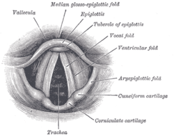
 Laryngoscopic view of the vocal folds. (Vestibular fold labeled at center right.) | |
 Cut through the larynx of a horse: 1 hyoid bone 2 epiglottis 3 vestibular fold, false vocal fold/cord, (Plica vestibularis) 4 vocal fold, true vocal fold, (Plica vocalis) 5 Musculus ventricularis 6 ventricle of larynx (Ventriculus laryngis) 7 Musculus vocalis 8 Adam's apple (thyroid cartilage) 9 rings of cartilage (cricoid cartilage) 10 Cavum infraglotticum 11 first tracheal cartilage 12 Windpipe (Trachea) | |
| Details | |
| Identifiers | |
| Latin | plica vestibularis, plica ventricularis |
| TA98 | A06.2.09.008 |
| TA2 | 3196 |
| FMA | 55452 |
| Anatomical terminology | |
The vestibular fold (ventricular fold, superior or false vocal cord) is one of two thick folds of mucous membrane, each enclosing a narrow band of fibrous tissue, the vestibular ligament, which is attached in front to the angle of the thyroid cartilage immediately below the attachment of the epiglottis, and behind to the antero-lateral surface of the arytenoid cartilage, a short distance above the vocal process.
The lower border of this ligament, enclosed in mucous membrane, forms a free crescentic margin, which constitutes the upper boundary of the ventricle of the larynx.
They are lined with respiratory epithelium, while true vocal cords have stratified squamous epithelium.
© MMXXIII Rich X Search. We shall prevail. All rights reserved. Rich X Search